File:Amphioxus Transverse sections through embryos of different ages.jpg
Original file (1,741 × 1,206 pixels, file size: 1.09 MB, MIME type: image/jpeg)
Captions
Captions
Summary
[edit]
| Description |
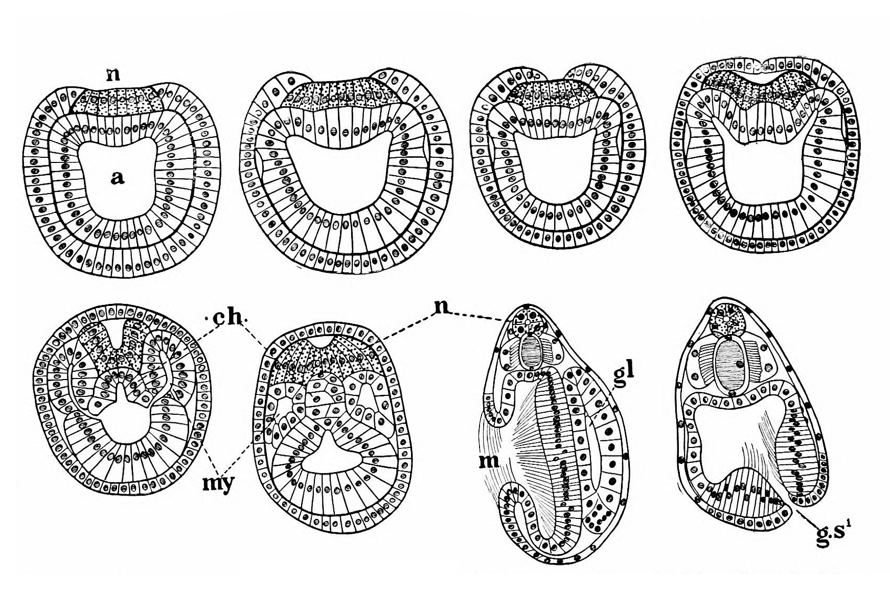
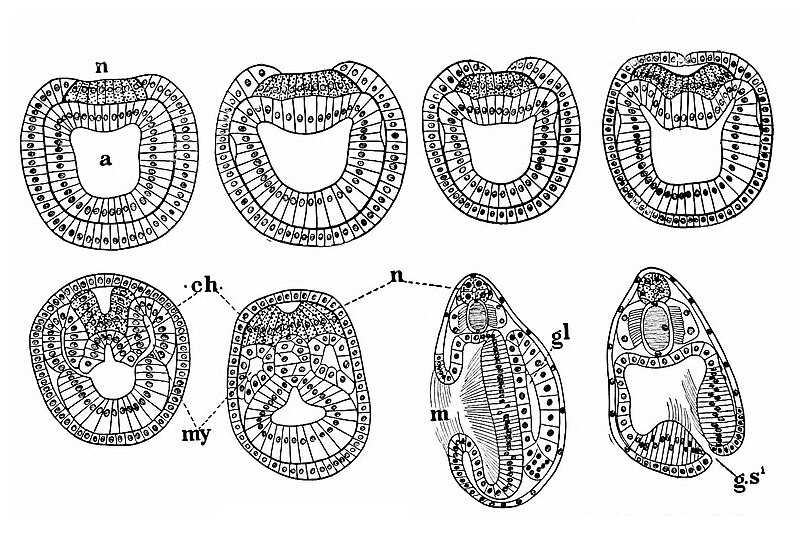
Fig. 65.— Transverse sections through embryos of different ages, illustrating the mode of formation and relations of the medullary tube, notochord, mesodermic somites, ete, (After HATSCHEK.) The figures should have Leen lettered consecutively from 1 to A (they are thus referred to in the text), but the letters were accidentally omitted. a, Archenteron.ch, Notochord. gl. Club-shaped gland, gs’. First primary gillslit, m, Mouth. my. Myoccelomic pouches (mesodermic somites). n. Medullary plate and tube, N.B.—In the last two figures the nuclei of the calomic epithelium, which has become extremely flattened, are not well indicated. ‘Ihe nuclei are scarce enough, but not quite so scarce as would appear from the figures. |
| Date | |
| Source | https://archive.org/details/cu31924001026131/page/119/mode/1up?view=theater Amphioxus and the ancestry of the vertebrates. New York, London, Macmillan. |
| Author | Willey, Arthur |
Licensing
[edit]
|
This work is in the public domain in its country of origin and other countries and areas where the copyright term is the author's life plus 70 years or fewer. | |
| This file has been identified as being free of known restrictions under copyright law, including all related and neighboring rights. | |

|
This file, which was originally posted to an external website, has not yet been reviewed by an administrator or reviewer to confirm that the above license is valid. See Category:License review needed for further instructions.
|
File history
Click on a date/time to view the file as it appeared at that time.
| Date/Time | Thumbnail | Dimensions | User | Comment | |
|---|---|---|---|---|---|
| current | 08:28, 24 March 2024 | | 1,741 × 1,206 (1.09 MB) | Rasbak (talk | contribs) | {{information |description=Fig. 65.— Transverse sections through embryos of different ages, illustrating the mode of formation and relations of the medullary tube, notochord, mesodermic somites, ete, (After HATSCHEK.) The figures should have Leen lettered consecutively from 1 to A (they are thus referred to in the text), but the letters were accidentally omitted. a, Archenteron.ch, Notochord. gl. Club-shaped gland, gs’. First primary gillslit, m, Mouth. my. Myoccelomic pouches (mesodermic so... |
You cannot overwrite this file.
File usage on Commons
There are no pages that use this file.