File:Amphioxus Growth of the ciliated embryo.jpg
Original file (1,735 × 2,138 pixels, file size: 1.26 MB, MIME type: image/jpeg)
Captions
Captions
Summary
[edit]
| Description |
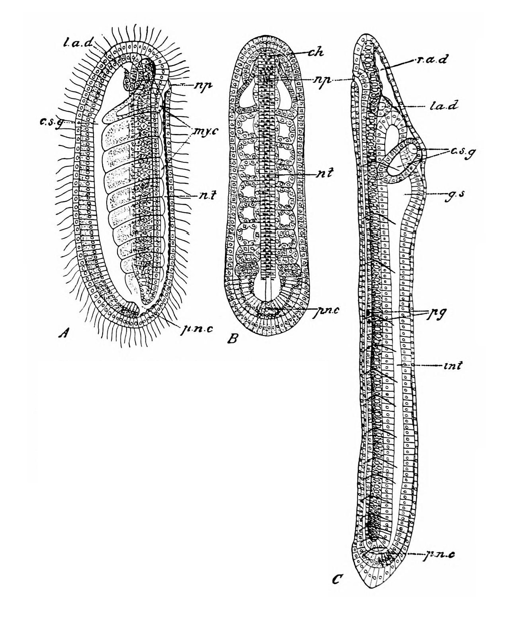
Fig. 63.— Growth of the ciliated embryo of Amphioxus. (After HATSCHEK, slightly altered.) B. Same stage from dorsal side. C. Stage, with fifteen pairs of myotomes; from the right side. Vacuoles have appeared in cells of notochord. ch. Notochord. c.s.g. Club-shaped gland. g.s. Rudiment of first gill-slit. int, Intestine. l.a.d. Left head-cavity (left anterior intestinal diverticulum). my.c. Myocoelomic (archenteron) pouches. np. Neuropore. n.t. Medullary tube. pg. Pigment granules in floor of medullary tube. p.n.c. Posterior neurenteric canal. r.a.d. Right head-cavity (right anterior intestinal diverticulum). |
| Date | |
| Source | https://archive.org/details/cu31924001026131/page/116/mode/1up?view=theater Amphioxus and the ancestry of the vertebrates. New York, London, Macmillan. |
| Author | Willey, Arthur |
Licensing
[edit]
|
This work is in the public domain in its country of origin and other countries and areas where the copyright term is the author's life plus 70 years or fewer.
| |
| This file has been identified as being free of known restrictions under copyright law, including all related and neighboring rights. | |

|
This file, which was originally posted to an external website, has not yet been reviewed by an administrator or reviewer to confirm that the above license is valid. See Category:License review needed for further instructions.
|
File history
Click on a date/time to view the file as it appeared at that time.
| Date/Time | Thumbnail | Dimensions | User | Comment | |
|---|---|---|---|---|---|
| current | 11:42, 24 March 2024 | | 1,735 × 2,138 (1.26 MB) | Rasbak (talk | contribs) | {{information |description=Fig. 63.— Growth of the ciliated embryo of Amphioxus. (After HATSCHEK, slightly altered.)<br> i. Stage, with nine pairs of myoccelomic pouches ; from left side. B. Same stage from dorsal side. C. Stage, with fifteen pairs of myotomes; from the right side. Vacuoles have appeared in cells of notochord. ch. Notochord. c.s.g. Club-shaped gland. g.s. Rudiment of first gill-slit. int, Intestine. l.a.d. Left head-cavity (left anterior intestinal diverticulum). my.c. Myo... |
You cannot overwrite this file.
File usage on Commons
There are no pages that use this file.