File:Alacaris multinoda fossil.jpg
Jump to navigation
Jump to search
Size of this preview: 569 × 600 pixels. Other resolutions: 228 × 240 pixels | 455 × 480 pixels | 729 × 768 pixels | 971 × 1,024 pixels | 1,998 × 2,106 pixels.
Original file (1,998 × 2,106 pixels, file size: 1,022 KB, MIME type: image/jpeg)
Captions
Captions
Add a one-line explanation of what this file represents
Summary
[edit]
| Description |
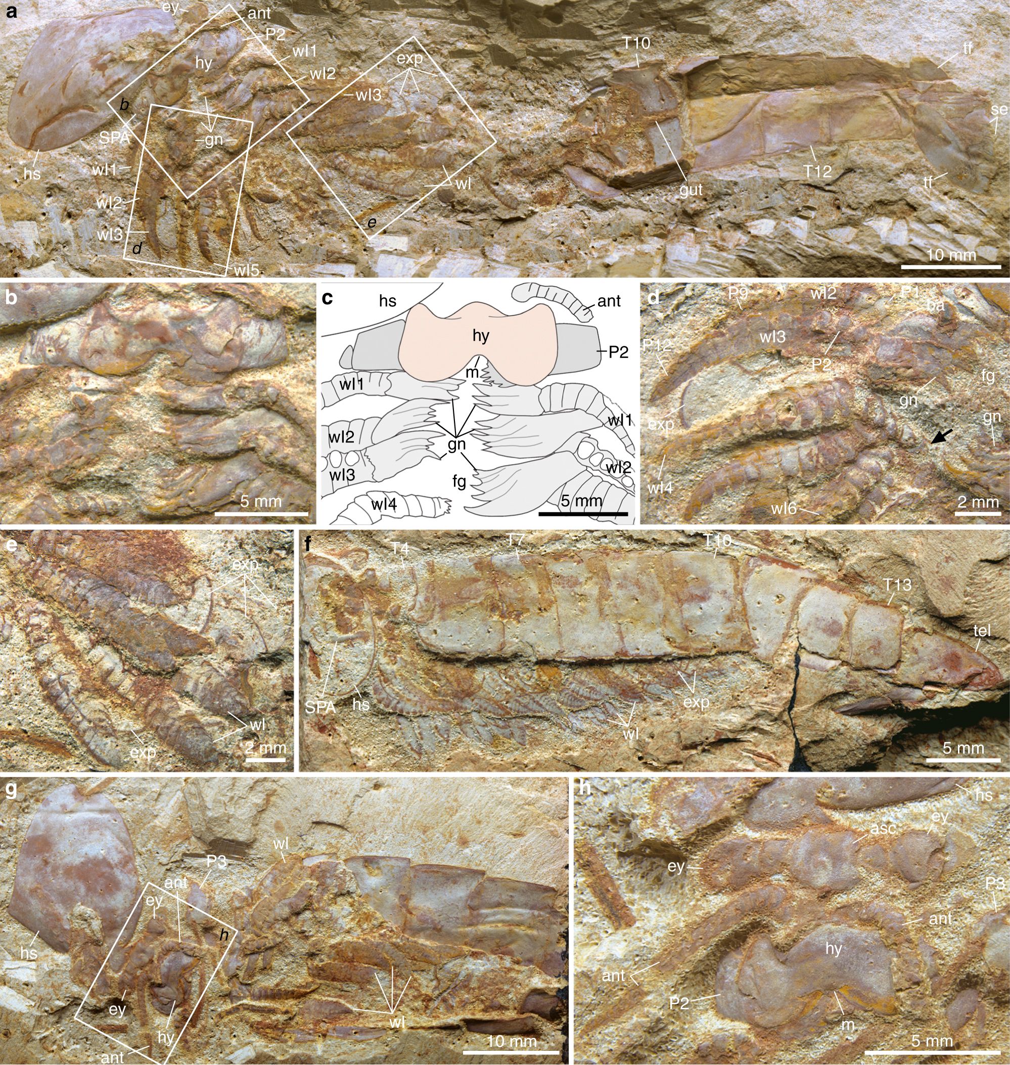
English: Alacaris multinoda from the Cambrian (Stage 3) Xiaoshiba Lagerstätte. a–e YKLP 12268 (holotype). a Complete individual in ventral view showing disarticulated head shield, appendicular organization, trunk tergites, and tailspine with paired flukes. b Details of area b, showing antennae, hypostome, SPAs, and three sets of walking legs with differentiated gnathobasic protopodites forming a ventral food groove. c Interpretative drawing of b. d Close-up of area d, showing multisegmented endopods with prominent protopodites, followed by walking legs with spinose endites (arrowed). e Close-up of area e, showing multisegmented endopods and flap-like exopods. f YKLP 12269, lateral view of a complete individual. g YKLP 12276, specimen with disarticulated head shield, showing organization of the anterior region. h Close-up of area h, showing the anterior sclerite with stalked eyes, the insertion of the paired antennae close to the anterior edge of the hypostome, and the proximal portions of the SPAs. ant: antenna, asc: anterior sclerite, exp: exopod, ey: eye, fg: food groove, gn: gnathobase, gut: alimentary canal, hs: head shield, hy: hypostome, m: mouth, Pn: podomeres, se: setae, SPA: specialized post-antennal appendage, tel: tailspine, tf: tail fluke, Tn: tegites, wln: walking legs |
| Date | |
| Source | Yang, J., Ortega-Hernández, J., Legg, D.A. et al. Early Cambrian fuxianhuiids from China reveal origin of the gnathobasic protopodite in euarthropods. Nat Commun 9, 470 (2018). https://doi.org/10.1038/s41467-017-02754-z |
| Author | Jie Yang, Javier Ortega-Hernández, David A. Legg, Tian Lan, Jin-bo Hou & Xi-guang Zhang |
| Permission (Reusing this file) |
This file is licensed under the Creative Commons Attribution 4.0 International license.
|
| Other versions |
.jpg)
File history
Click on a date/time to view the file as it appeared at that time.
| Date/Time | Thumbnail | Dimensions | User | Comment | |
|---|---|---|---|---|---|
| current | 13:59, 8 April 2020 | | 1,998 × 2,106 (1,022 KB) | Hemiauchenia (talk | contribs) | Transferred from https://media.springernature.com/full/springer-static/image/art%3A10.1038%2Fs41467-017-02754-z/MediaObjects/41467_2017_2754_Fig1_HTML.jpg |
You cannot overwrite this file.
File usage on Commons
The following page uses this file:
File usage on other wikis
The following other wikis use this file: